Posterior Cervical Foraminotomy
A minimally invasive procedure that enlarges the neural foramen (the space where nerves exit the spine) by removing small amounts of bone from the back of the neck. Ideal when nerve compression is localized and fusion is not needed.
What is Posterior Cervical Foraminotomy?
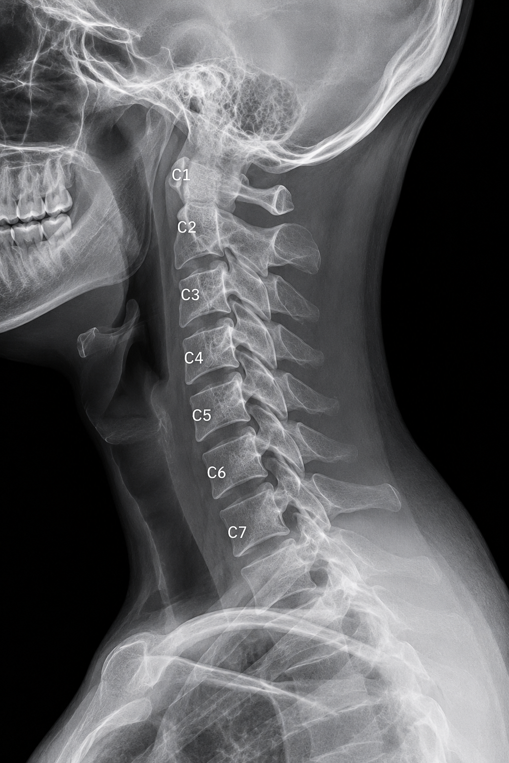
Posterior cervical foraminotomy is a surgical procedure that enlarges the neural foramen (the opening through which nerve roots exit the spine) by removing a small amount of bone from the back of the neck.
The Concept
When a lateral (side) disc herniation or bone spur narrows the neural foramen and presses on an exiting nerve root, this procedure widens the opening to relieve the compression. Unlike disc removal procedures, the disc itself remains intact.
When Indicated
- Lateral disc herniation compressing a nerve root
- Foraminal bone spur (osteophyte) narrowing nerve passage
- Single-level nerve compression without central cord involvement
- Radiculopathy (arm pain/numbness) as primary symptom
Patient Selection
- No significant instability at the affected level
- Acceptable surgical candidate in reasonable health
- Failed conservative management or progressive neurological deficit
- Clear imaging correlation between pathology and symptoms
The Posterior Approach & Anatomy
This procedure uses a posterior (back-of-neck) approach with precise anatomical technique to access and enlarge the neural foramen.
Posterior Access and Key Structures
The surgeon works from the back of the neck, using specialized microscopic visualization and instruments to safely enlarge the narrow passage where the nerve exits the spine.
Anatomical Landmarks
- Incision location — midline or paramedian incision on the back of the neck over the affected level
- Muscle reflection — paraspinal muscles are gently moved aside to expose the lamina and facet joints
- Neural foramen — bounded by bone above and below, and the facet joint towards the spine
- Microscopic technique — high magnification allows precise bone removal without damaging the nerve
The Surgical Procedure
Posterior cervical foraminotomy is performed under microscopic visualization with meticulous attention to nerve protection.
-
Positioning and exposure
You are positioned prone (face down). A small midline or paramedian incision is made over the affected level. The surgeon carefully reflects the paraspinal muscles to expose the lamina (bony arch) and facet joint.
-
Microscopic visualization
An operating microscope is used to visualize the neural foramen, allowing precise identification of the compressed nerve root and surrounding bone landmarks.
-
Partial laminectomy
A small portion of the lamina is carefully removed, widening the opening on the upper border of the foramen. This is performed with fine instruments and constant attention to nerve protection.
-
Medial facetectomy
The inner (medial) portion of the facet joint is carefully removed to further enlarge the foramen. The disc remains intact, and no fusion or instrumentation is needed.
-
Nerve decompression
Once the foramen is widened, the compressed nerve root is free of pressure. The surgical field is irrigated, hemostasis is achieved, and the muscles and skin are closed in layers.
Benefits of This Minimally Invasive Approach
Posterior cervical foraminotomy offers distinct advantages for appropriate patients.
Structural Advantages
- Preserves all anterior spinal structures
- No removal of disc material
- No hardware or fusion needed
- Maintains full segmental motion
Recovery & Lifestyle
- Faster recovery with minimal muscle disruption
- No fusion time required — immediate stability
- No long-term restrictions on neck motion
- Often effective for radicular (arm) pain relief
This is a nerve-root focused procedure. When nerve compression is strictly localized to the exit channel (neural foramen), foraminotomy can be highly effective while preserving maximum spinal architecture and function.
Common Questions
Questions patients often ask about posterior cervical foraminotomy.
Is this less invasive than other neck surgeries?+
Yes, in several ways. The procedure removes minimal bone and no disc material, requires no fusion or hardware, and preserves full motion. The posterior approach disrupts fewer anterior structures. However, "minimally invasive" is relative — it is still an operative procedure performed under general anesthesia.
What is the recovery timeline?+
Initial healing takes 4–6 weeks. Return to desk work may occur within 2–4 weeks. Progressive return to activity typically occurs over 2–3 months. Your surgeon will provide specific guidelines based on your individual healing.
Will arm pain improve right away?+
Many patients experience significant improvement in arm pain and numbness within weeks as the nerve is decompressed. However, complete symptom resolution may take several weeks as nerve inflammation subsides and healing progresses.
Are there any restrictions afterward?+
In the first 6 weeks, avoid heavy lifting and strenuous activity. After healing, there are no permanent restrictions. The procedure preserves motion, so normal neck movement and activity are encouraged once cleared by your surgeon.
What happens if the nerve compression comes back?+
Recurrence at the same level is uncommon. If it occurs, revision foraminotomy or another procedure may be considered. Discuss long-term outcomes and the small risk of recurrence with your surgeon before surgery.
Want to know more?
Discuss with your surgeon whether posterior foraminotomy is an appropriate option for your condition.